 Lumbar Spine Anatomy: Overview, Gross Anatomy, Natural Variants
Lumbar Vertebrae - Anatomy Pictures and Information Can Be Fun For Anyone

Together, they improve the protection of the spinal cord and roots. The dura is the most shallow however resilient layer. The pia and arachnoid, together described the leptomeninges, are frail. The spine, roots, and nerve rootlets are closely invested by the pia. The dura and arachnoid together form a loose sheath (described dural/thecal sac) around these structures, separated from the canal walls by the epidural space.
The external surface area is rough and mixes with loose connective tissue in the epidural area. The internal surface area, facing into the subdural space, is smooth and covered by a layer of mesothelium. Inferiorly, the dural sac ends at the sacral canal, normally at S2-S3 (in some cases S1). The dura continues caudally as a fibrous thread named the filum terminale externum or coccygeal ligament, which blends with the PLL over the coccyx.
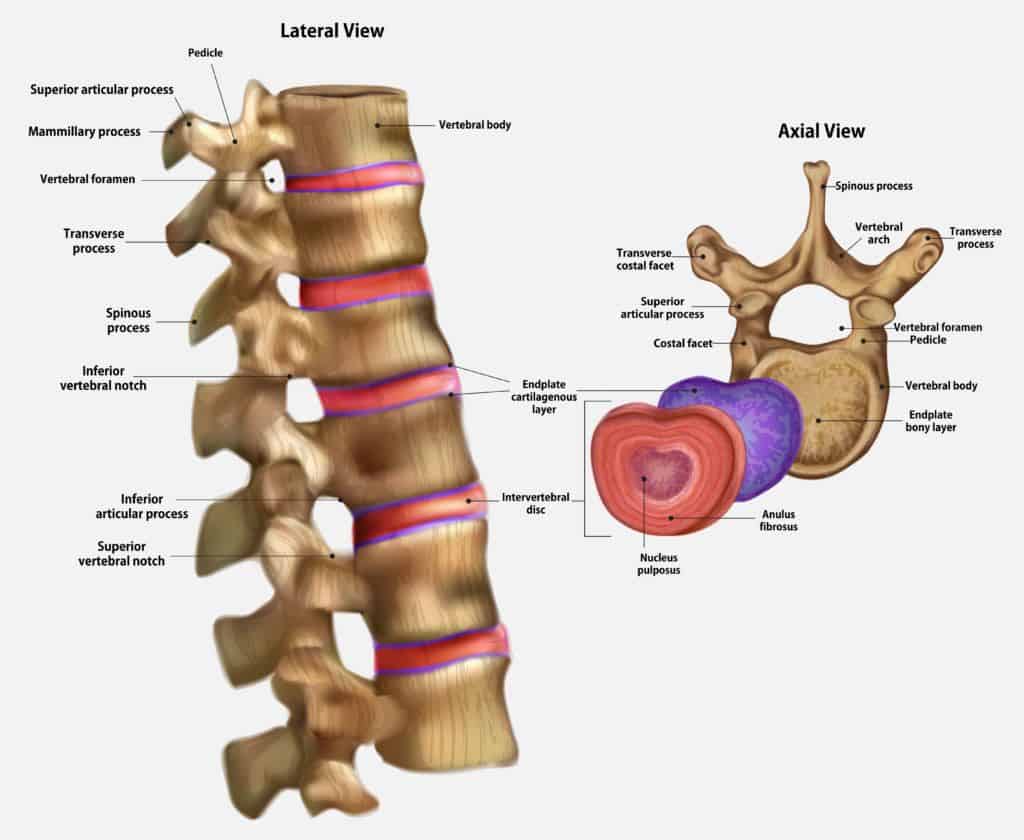
 Lumbar Spine Injury Illustration - High Impact® Visual Litigation Strategies™
Get This Report on Nomenclature and Classification of Lumbar Disc Pathology
Connective tissue insinuates the foramen anchor the dural sleeves so that they can protect the spine nerve roots from being stretched during L-spine movements. In addition to these tetherings, the dura is connected in locations to the PLL. Epidural space The epidural (peridural/extradural) area ends inferiorly at the sacral hiatus, where it is sealed by the posterior sacrococcygeal ligaments.
The entire area is occupied by loose connective tissue with variable fat content, providing padding around the dural sac and spine and functioning as a type to hold the thin internal vertebral plexus of veins open. The vertebral venous plexus is embedded in the epidural loose connective tissue, often transmitting big amounts of blood.
Getting The Lumbar Spine Anatomy -Redlands, Loma Linda, Highland To Work
A layer of mesothelium covers all leptomeningeal surface areas bathed by cerebrospinal fluid (CSF). The arachnoid mater lines the entire dural sac and extends into the dural sleeves. It likewise sends trabeculae throughout the subarachnoid space to the pia, facilitating CSF blending. Along the posterior midline, the trabeculae form a well-defined subarachnoid septum.
The pia mater offers support for the vasculature and nerves in the subarachnoid space. Source adheres totally to the spine cord. The pia forms a different sheath for each nerve rootlet and root as far laterally as the foramen, blending with the epineurium. Caudally, the pia continues as the thin filum terminale internum.


 icons at the top right corner of the subsection.